
Open Journal of
Clinical and Medical Images
Case Report - Open Access, Volume 4
Breathlessness and T-wave inversion: Uncovering perfusion defects in apical hypertrophic cardiomyopathy
Simran Shergill*; Gerry P McCann; Jayanth Ranjit Arnold
Department of Cardiovascular Sciences, University of Leicester, Glenfield Hospital, Leicester, United Kingdom.
*Corresponding Author: Simran Shergill
Department of Cardiovascular Sciences, University of
Leicester, Glenfield Hospital, Leicester, United Kingdom.
Email: Simran.shergill@nhs.net
Received : Dec 19, 2023
Accepted : Jan 12, 2024
Published : Jan 18, 2024
Archived : www.jclinmedimages.org
Copyright : © Shergill S (2024).
Abstract
A 72-year-old female presents with breathlessness. ECG demonstrated deep T-wave inversion in the praecordial leads. Echocardiogram was limited but demonstrated no obvious impairment in left ventricular systolic function or significant valve dysfunction. CMR was conclusive for a diagnosis of apical hypertrophic cardiomyopathy with additional microvascular dysfunction in the thickened and dysfunctional apical segments. This case highlights the uniquely powerful nature of CMR in its ability to accurately visualise and quantify ventricular morphology, with the additional strengths to characterise myocardial tissue and the non-invasive assessment of microvascular dysfunction.
Keywords: Apical hypertrophic cardiomyopathy; Left ventricular hypertrophy; Cardiovascular magnetic resonance; Stress perfusion MRI; Microvascular dysfunction.
Citation: Shergill S, McCann GP, Arnold JR. Breathlessness and T-wave inversion: Uncovering perfusion defects in apical hypertrophic cardiomyopathy. Open J Clin Med Images. 2024; 4(1): 1162.
Background
A 72-year-old female of South Asian descent presented to the cardiology clinic with increasing breathlessness and effort intolerance. There were no associated symptoms of angina, orthopnoea or peripheral oedema. Past medical history included a stroke several years previously with no residual limiting neurological deficit. On assessment, she was normotensive (blood pressure 120/75 mmHg) with no audible murmurs or features of congestive cardiac failure. Resting 12-lead electrocardiogram (ECG) demonstrated normal sinus rhythm with voltage criteria for left ventricular hypertrophy (LVH) and deep anterolateral Twave inversion. There was no accompanying history of uncontrolled hypertension or relevant family history. A transthoracic echocardiogram was limited but demonstrated no obvious impairment in LV systolic function or significant valve dysfunction. However, all myocardial segments were not clearly visualised.
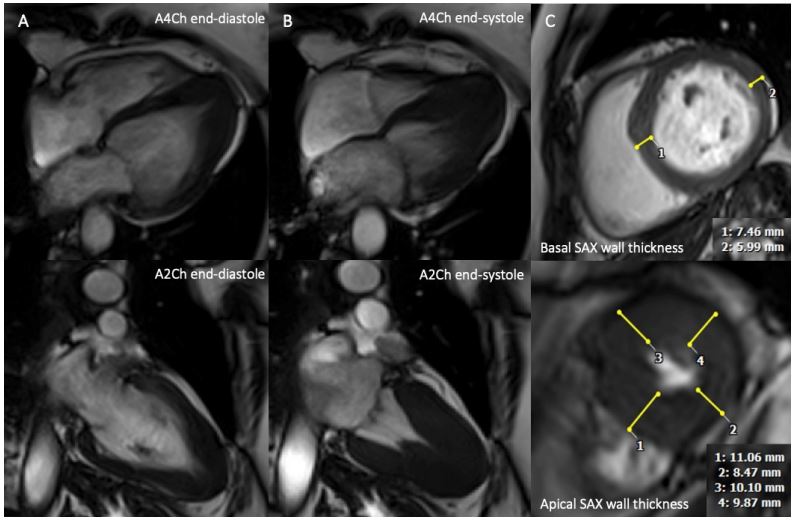
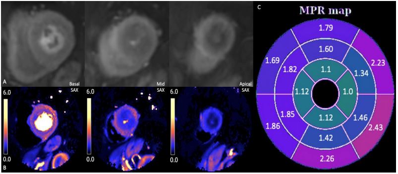
CMR examination: Cardiac magnetic resonance (CMR) (3T scanner, MAGNETOM Vida, Siemens) steady-state free precession (SFFP) cine images demonstrated normal LV volumes and systolic function but a lack of tapering in wall thickness towards the apex, with associated apical systolic cavity obliteration (Figure 1A and B). There was significant relative hypertrophy of the apical segments (maximally 11 mm vs. 7 mm basally) with a calculated apical to posterior wall thickness ratio of 1.8 (Figure 1C). Native T1-mapping using a modified Look-Locker (MOLLI) sequence revealed moderately elevated myocardial T1 times towards the apex (1300-1350 ms vs. 1250 ms basally) (Figure 2A) and resultant normal extracellular volume fraction (ECVF). Postcontrast Look-Locker TI scout demonstrated normal myocardial nulling kinetics and late gadolinium enhancement (LGE) imaging utilising magnitude and phase-sensitive inversion recovery (PSIR) sequences demonstrated low-grade diffuse fibrosis in the affected apical segments (Figure 2B and C). Qualitative and automated inline quantitative myocardial perfusion mapping demonstrated a prominent circumferential perfusion defect at the mid-apical levels (Figure 3A and B), which was confirmed on quantitative perfusion analysis with a significant reduction in myocardial perfusion reserve (MPR) towards the apex (1-1.1 vs. 1.8-2.2 basally) (Figure 3C). The perfusion defect was more pronounced in the apical endocardial layers during stress with a subsequent MPR gradient between endocardial and epicardial layers of ~-50-60%. The CMR findings were conclusive for a diagnosis of relative apical hypertrophic cardiomyopathy (ApHCM) with associated low-grade fibrosis and microvascular dysfunction in the affected hypertrophied segments.

Discussion
This case highlights the uniquely powerful nature of CMR in its ability to accurately visualise and quantify ventricular morphology, with the additional strengths to characterise myocardial tissue and the non-invasive assessment of microvascular dysfunction, both of which are of paramount importance in assessing patients with increased wall thickness. We were able to confidently confirm the presence of sarcomeric ApHCM given the moderate elevations in T1 times, normal myocardial nulling kinetics and ECVF, which excluded phenocopies such as Anderson-Fabry disease and cardiac amyloidosis. Current European Society of Cardiology guidance recommends the diagnosis of HCM be made with increased wall thickness of ≥15 mm in ≥1 myocardial segment in the absence of loading conditions [1]. Often the diagnosis of ApHCM fails to meet this criterion (the so-called “relative” ApHCM picture)., However, the presence of deep praecordial T-wave inversion on ECG, lack of wall thickness tapering towards the apex [2] and apical to posterior wall thickness ratio of ≥1.5 [3] supports the diagnosis. During stress perfusion imaging, a circumferential perfusion defect was observed, affecting the thickened segments in the mid-apical LV cavity with a further reduction in blood flow in the subendocardial layer. Perfusion defects have been highlighted as a universal feature in ApHCM [4] and likely to be multifactorial due to a combination of a coronary steal phenomenon, diastolic contractile persistence [5], myocyte disarray and mechanical compression from the hypertrophied and hyperdynamic apical segments.
References
- Arbelo E, Protonotarios A, Gimeno JR, Arbustini E, Barriales-Villa R, Basso C et al. 2023 ESC Guidelines for the management of cardiomyopathies: Developed by the task force on the management of cardiomyopathies of the European Society of Cardiology (ESC). European heart journal. 2023; 44(37): 3503-626.
- Flett AS, Maestrini V, Milliken D, Fontana M, Treibel TA, Harb R et al. Diagnosis of apical hypertrophic cardiomyopathy: T-wave inversion and relative but not absolute apical left ventricular hypertrophy. International Journal of Cardiology. 2015; 183: 143-8.
- Eriksson MJ, Sonnenberg B, Woo A, Rakowski P, Parker TG, Wigle ED et al. Long-term outcome in patients with apical hypertrophic cardiomyopathy. Journal of the American College of Cardiology. 2002; 39(4): 638-45.
- Hughes RK, Augusto JB, Knott K, Davies R, Shiwani H, Seraphim A et al. Apical Ischemia Is a Universal Feature of Apical Hypertrophic Cardiomyopathy. Circulation: Cardiovascular Imaging. 2023; 16(3): 014907.
- Stephenson E, Monney P, Pugliese F, Malcolmson J, Petersen SE, Knight C et al. Ineffective and prolonged apical contraction is associated with chest pain and ischaemia in apical hypertrophic cardiomyopathy. Int J Cardiol. 2018; 251: 65-70. doi: 10.1016/j. ijcard.2017.09.206.