
Open Journal of
Clinical and Medical Images
Case Report - Open Access, Volume 4
Atypical presentation of pulmonary embolism masquerading as abdominal pain
*Corresponding Author: Hasan Hazim Alsararatee
Acute Medicine/SDEC, Northampton General Hospital
NHS Trust, Northampton, Northampton shire, UK.
Email: hasan.alsararatee@nhs.net
Received : Jan 23, 2024
Accepted : Feb 19, 2024
Published : Feb 26, 2024
Archived : www.jclinmedimages.org
Copyright : © Alsararatee HH (2024).
Abstract
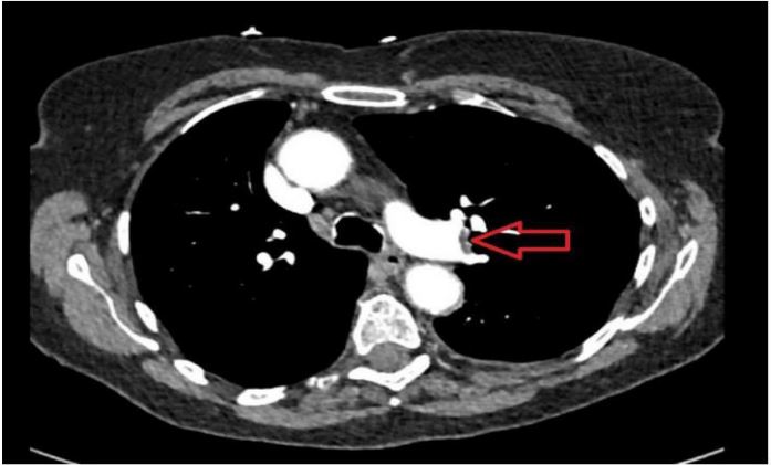
This case report highlights a rare presentation of Pulmonary Embolism (PE) with atypical abdominal pain, emphasizing the challenges in timely diagnosis. A female patient in her early eighties presented with left abdomen pain, initially treated for Urinary Tract Infection (UTI), and later accurately diagnosed with PE through Computed Tomography Pulmonary Angiography (CTPA). The mechanisms of abdominal pain encompass many hypotheses such as right-sided heart failure-induced hepatic congestion, diaphragmatic irritation from the pulmonary lobe infraction, increased blood viscosity, and pulmonary hypertension-associated abdominal lymphedema. PE is a prominent cause of unexpected deaths in hospitalized patients, with misdiagnosis occurring in up to 70% of cases, often only discovered postmortem. The condition manifests through various symptoms, with dyspnoea and chest pain being the most common. Abdominal pain, though less frequent, has been documented with an incidence of 6.7% in previous studies.
Citation: Alsararatee HH. Atypical presentation of pulmonary embolism masquerading as abdominal pain. Open J Clin Med Images. 2024; 4(1): 1167.
Background
PE is one of the comments presentations to Emergency Department (ED) and often is misdiagnosed leading to high mortality and morbidity [1]. The missed diagnosis rate for PE reportedly exceeds 70%, with only 7% of deceased PE patients diagnosed before death [2]. In contrast, while the hospitalization mortality rate for PE ranges from 2.5% to 10%, the mortality rate for those with a missed diagnosis is typically acknowledged to be 30% [3].
However, some literature highlights that the actual prevalence of PE remains uncertain. Inaccurate estimates of PE prevalence may, to some extent, stem from the under recognition of atypical presentations of this condition [4]. Abdominal pain emerges as an atypical manifestation of Pulmonary Embolism [PE], with a notable association highlighted in a comprehensive 2011 study where 10.7% of 1880 diagnosed PE patients reported upper abdominal pain [5]. Additionally, rare instances of resolved abdominal pain following anticoagulant treatment have been reported as part of these atypical presentations [6]. Thus, Physicians must uphold a systemic approach and recognize the imperative need to contemplate the potentiality of a fatal diagnosis, particularly in cases characterized by unexplained abdominal pain.
Case presentation
A female patient in her early eighties was referred from ED to Same Day Emergency Care (SDEC) due to the sudden onset of left sharp abdominal pain persisting for 72 hours. With a medical history encompassing hypertension and hypercholesterolemia, she denied any dysuria or haematuria. No recent trauma or falls, or any association with COVID-19 or COVID-19 vaccinations. Additionally, there was no significant family history of respiratory or cardiovascular diseases. Initial assessments by surgical and urology teams concluded that acute surgical intervention was unnecessary, and an abdominal ultrasound revealed no abnormalities. However, a urine culture indicated White Blood Cells (WBC) >200 and urinalysis (Dipstick) shows leukocytes of ++, and Nitrates of ++, prompting treatment for a Urinary Tract Infection (UTI). During examination, blood pressure measured 132/80 mmHg, and the heart rate was 75 beats per minute. She was a well-nourished female, slightly uncomfortable in bed, without jugular Venus distention or calf tenderness/erythema. There were no murmurs or rubs but fine crackles in the left lower base. The abdomen was tender in the left upper quadrant, with no signs of rebound or guarding. Murphy’s sign was negative. No lower limbs pitting oedema. The Wells score indicated a moderate risk (3), and the D- dimer level measured 2210 ng/m, leading to a referral to medical SDEC for a review of possible PE.
Investigations
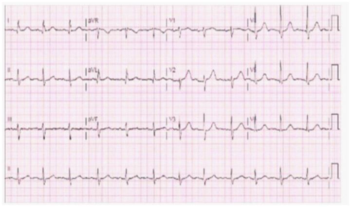
Blood investigations revealed a C-Reactive Protein CRP level of 114 mg/L (normal range: 0-5) and a WBC of 14.0x10⁹ /L (normal range: 4.0-10.0). Troponin T levels were below 13 ng/L, and urea and electrolytes remained within normal limits. The D-dimer test indicated elevated levels at 2210 ng/m. A COVID-19 test yielded negative results, and blood cultures along with sputum gram stain with culture returned negative results. The Electrocardiogram (ECG) displayed a normal sinus rhythm (Figure 1).
Treatment
She was treated for UTI with oral Fosfomycin 3 g and treated for unprovoked PE with Rivaroxaban 15 mg BD for 21 days followed by 20 mg OD for 4 months. The patient was covered with oral Co-amoxiclav 625 mg TDS for 5 days for community acquired pneumonia as well since developing cough with high inflammatory markers. After five days, the cough and abdominal pain are completely resolved.
Outcome and follow-up
After five days of completing the above antibiotics, the cough and abdominal pain are completely resolved. She is doing well on four weeks follow up.
Discussion
The clinical manifestations of PE are often nonspecific, posing challenges to diagnosis. Notably, acute surgical abdomen is not commonly listed as presenting features of PE. The typical presentation of PE encompasses dyspnoea (80%), chest pain (52%), cough (20%), haemoptysis (11%), and syncope (19%) [7]. Conversely, the atypical presentation includes abdominal pain, high fever, new-onset atrial fibrillation, and disseminated intravascular coagulation [7]. When abdominal pain is present in the context of PE, it is usually accompanied by other typical PE symptoms, highlighting the importance of recognizing the diverse clinical manifestations of this condition. However, this was not the case in our case study as the patient has left upper quadrant and flank pain.
When patients present with abdominal pain, healthcare professionals prioritize abdominal pathologies, considering myocardial infarction or pleurisy, rarely considering pulmonary embolism [8]. In this case report, the patient initially attended with abdominal pain without typical PE symptoms such as pleuritic chest pain or shortness of breath, was initially misdiagnosed with UTI. In our case report, it was only after observing an elevated D-dimer level and conducting a comprehensive examination the suspicion of PE emerged.
A variety of potential mechanisms that contribute to abdominal pain associated with PE are explained. One perspective suggests that PE can induce abdominal pain, attributing the pain to hepatic congestion resulting from right-sided heart failure induced by PE [9]. Previous literature supports that this explanation is considered a primary cause of PE-related abdominal pain [10]. However, this was not the case with our case report as the patient did not have RV dilation. Increased right ventricle pressure leads to paradoxical embolism and reduced blood supply to abdominal organs [10]. Another theory suggests that increasing the blood viscosity, alongside low oxygen levels, may generate small emboli causing focal necrosis in abdominal organs which leads to abdominal pain [11]. However, another theory explains that pulmonary hypertension associated with PE is implicated in abdominal lymphedema and hepatobiliary portal infiltration [12]. Additionally, previous literature points out that abdominal pain may be referred to the chest and causing chest pain, potentially arising from pulmonary hypertension or thrombus stimulation of sensory nerve endings in blood vessel walls, or lateral diaphragmatic stimulation [12]. Alternatively, abdominal pain may result from diaphragmatic irritation from the pulmonary lobe infraction. In our case report, the abdominal pain might be from the infraction of pneumonia [13].
In conclusion, this case highlights a significant example of PE that could have been easily missed without a heightened level of suspicion. It emphasizes the critical need for a comprehensive and systematic approach when investigating nonspecific symptoms, emphasizing that abdominal pain can serve as the primary and sole presenting complaint in PE, leading to a diagnosis that can be lifesaving. Recognizing abdominal and flank pain as potential presenting symptoms of PE is essential to prevent diagnostic delays. It underscores the imperative for clinicians to remain vigilant, as a missed diagnosis of this potentially devastating condition carries significant consequences.
Learning points/Take-home messages
- PE should be considered as a differential diagnosis even in the absence of typical respiratory or cardiac symptoms such as breathlessness, tachycardia, chest pain, haemoptysis, and syncope.
- Abdominal pain can be atypical presentation of PE and therefore clinicians should assess and examine patients comprehensively.
- Abdominal pain in PE can be result from diaphragmatic irritation from the pulmonary infarction lobe, hepatic congestion, paradoxical embolism and reduced blood supply to abdominal organs. Increased blood viscosity, abdominal lymphoedema from the pulmonary hypertension or stimulation of sensory nerves.
Patients’ perspective
The patient expresses heartfelt gratitude for the attentive care and the discovery of Pulmonary Embolism (PE) through the CT scan. She is genuinely thankful for the dedicated medical teams and the author for their commitment to her well-being.
References
- Cooper J. Improving the diagnosis of pulmonary embolism in the emergency department. BMJ quality improvement reports. 2015; 4(1): u208698w4222. https://doi.org/10.1136/bmjquality.u208698.w4222
- Cohen AT, Agnelli, G, Anderson FA, Arcelus JI, Bergqvist D, et al. VTE Impact Assessment Group in Europe (VITAE). Venous thromboembolism (VTE) in Europe. The number of VTE events and associated morbidity and mortality. Thrombosis and haemostasis. 2007; 98(4): 756-764.
- Konstantinides SV, Barco S, Lankeit M, Meyer G. Management of Pulmonary Embolism: An Update. Journal of the American College of Cardiology. 2016; 67(8): 976-990. https://doi.org/10.1016/j.jacc.2015.11.061
- Jolobe OPM. Atypical manifestations of pulmonary embolism. Arch Vas Med. 2020; 4: 008-018.
- Fields JM, Dean AJ. Systemic causes of abdominal pain. Emergency medicine clinics of North America. 2011; 29(2): 195-vii. https://doi.org/10.1016/j.emc.2011.01.011
- Carroll BJ, Heidinger BH, Dabreo DC, Matos JD, Mohebali D, et al. Multimodality Assessment of Right Ventricular Strain in Patients With Acute Pulmonary Embolism. The American journal of cardiology. 2018; 122(1): 175-181. https://doi.org/10.1016/j.amjcard.2018.03.013
- Han Y, Gong Y. Pulmonary embolism with abdominal pain as the chief complaint: A case report and literature review. Medicine. 2019; 98(44): e17791. https://doi.org/10.1097/MD.0000000000017791
- Govender I, Rangiah S, Bongongo T, Mahuma P. A Primary Care Approach to Abdominal Pain in Adults. South African family practice: Official journal of the South African Academy of Family Practice/Primary Care. 2021; 63(1): e1-e5. https://doi.org/10.4102/safp.v63i1.5280
- Rehman H, John E, Parikh P. Pulmonary Embolism Presenting as Abdominal Pain: An Atypical Presentation of a Common Diagnosis. Case reports in emergency medicine. 2016; 2016: 7832895. https://doi.org/10.1155/2016/7832895
- Vyas V, Goyal A. Acute Pulmonary Embolism. In StatPearls. StatPearls Publishing. 2022.
- Mahabir VSD, Hosein AS, Giddings SL. Response to ‘Pulmonary embolism: an often forgotten differential diagnosis for abdominal pain’. QJM: Monthly journal of the Association of Physicians. 2020; 113(1): 70. https://doi.org/10.1093/qjmed/hcz183
- Gantner J, Keffeler JE, Derr C. Pulmonary embolism: An abdominal pain masquerader. Journal of emergencies, trauma, and shock. 2013; 6(4): 280-282. https://doi.org/10.4103/0974-2700.120376
- Al-Mane N, Al-Mane F, Abdalla Z, McDonnell J. Acute Surgical Abdomen: An Unusual Presentation of Pulmonary Embolus. Journal of investigative medicine high impact case reports. 2014; 2(3): 2324709614542339. https://doi.org/10.1177/2324709614542339.