
Open Journal of
Clinical and Medical Images
Case Report - Open Access, Volume 4
Pelvic radiation-induced sarcoma following treatment of cervical cancer
Guansheng Chen; Yongjun Wang; Lingyu Liu; Wenjing Li*
Department of Gynecology and Obstetric, Beijing Jishuitan Hospital, Capital Medical University, Beijing, China.
*Corresponding Author: Wenjing Li
Department of Gynecology and Obstetric, Beijing Jishuitan Hospital, Capital Medical University, Beijing, China.
Tel: +86-010-58398060 & +86-010-58398060;
Email: 38982606@qq.comReceived : Jan 24, 2024
Accepted : Feb 21, 2024
Published : Feb 28, 2024
Archived : www.jclinmedimages.org
Copyright : © Li W (2024).
Abstract
Post-radiation sarcomas account for 0.5-5.5% of all sarcomas, and include extra skeletal osteosarcoma, fifibrosarcoma leiomyosarcoma, and malignant fifibrous histiocytoma. This article presents a case of a 53-year-old female who developed a mixed-type postradiation sarcoma three years after radiotherapy for cervical cancer. Unfortunately, the patient succumbed to the condition six months after undergoing pelvic clearance surgery. No prior relevant case reports have been identified in the literature. Consequently, we have systematically reviewed and summarized this case, conducting a comprehensive literature review to contribute valuable insights into the understanding of post-radiation sarcomas. Our objective is to underscore the importance of vigilance regarding these conditions and to provide clinicians with pertinent clinical data for informed decision-making in the realm of tumor radiotherapy.
Keywords: Post-radiation sarcomas; Cervical cancer; Radiation; Treatment.
Abbreviations: RIS: Radiation Induced Sarcomas; PRS: Post-Radiation Sarcomas; RT: Radiotherapy; MFH: Malignant Fibrous Histiocytomas; RIAS: Radiation-Induced Angiosarcoma; CAC: Cervical Adenocarcinoma; MPNST: Malignant Peripheral Nerve Sheath Tumor; CCRT: Concurrent Chemo radiotherapy.
Citation: Chen G, Wang Y, Liu Y, Li W. Pelvic radiation-induced sarcoma following treatment of cervical cancer. Open J Clin Med Images. 2024; 4(1): 1168.
Introduction
Radiation induced Sarcomas (RIS) known as Post-Radiation Sarcomas (PRS) are rare long-term complications of Radiotherapy (RT), accounting for 0.5-5.5% of all sarcomas [1,2] developing within the radiation field or on its border. PRS histologic types included osteosarcoma, leiomyosarcoma, angiosarcoma, Schwannosarcoma, Malignant Fibrous Histiocytomas (MFH), and undifferentiated or clear cell sarcoma [3,4]. Histological confirmation of sarcoma prior history of RT, latency periods of several years, development of sarcoma within a previously irradiated field. We had to consider the diagnosis of PRS when patients meet the following condition: 1) prior history of RT, 2) development of sarcoma within a previously irradiated field, 3) latency periods of several years, 4) different pathology type from the original tumor. PRS appear to be resistant to further RT/chemotherapy. Radical surgery is the only treatment to improve disease-free survival. Tumors that are superficial and are 5 cm or smaller, regardless of grade, are likely to have a favorable prognosis and are therefore treated with a margin-negative excision [5]. Radiotherapy used to treat invasive breast tumors is a known risk factor for the development of the so-called Radiation-Induced Angiosarcoma (RIAS). Treatment is mostly surgical, and mastectomy with negative margins is considered the standard procedure [6].
RT is the main treatment of the cervical cancer stage IIB-IV. To date, the reports of sarcoma secondary to cervical cancer were rare. The time interval between the development of sarcoma after radiotherapy was relatively long usually more than five years [4]. Herein, we report a case of a 53-year-old female with RIS with rhabdomyoblastic differentiation of the cervix cancer. To be surprised, the onset of RIS happend quickly after 3 years of RT. Subsequently, we shall provide a comprehensive exposition of the patient’s overall diagnostic and therapeutic journey.
Case presentation
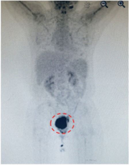
A 53-years-old female presented with a rapidly progressive hard mass on vagina, for the past 3 months accompanied by foul smelling, discontinuous minor bleeding. On examination, the mass measured 7.0x5.0x3.0 cm located in vagina residue, excentric growth and adhere to anterior vaginal wall, white surface and the mass surface was ulcerated. No local lymphadenopathy was found. Computed-tomography imaging revealed a soft tissue mass in pelvic, close relations with urethra, PET/ CT revealed the mass have a high metabolic activity (SUVmax 8.6), 4.4x5.7x7.4 cm and the boundary with bladder was not clear (Figure 1). Tumor markers was normal. Cytoscope showed the mucous of bladder’s trigone was pale and stiff. Clinicoradiological diagnosis was suggestive of post-radiation sarcomas. She was a follow up case of Cervical Adenocarcinoma (CAC) stage IIB 3 years ago and had tumor excision, following radiotherapy (DT 48.6Gy/1.8Gy/27fx, combined with intracavitary afterloading therapy 42Gy/6Gy/7fx). Reexamination after radiotherapy six months and 12 months later was normal. Unluckily, she suffered poorly differentiated adenocarcinorma of endometrium, performed hysterectomy, bilateral oophorosalpingectomy, omentum resection and appendectomy and followed six times TC chemotherapy.
Therapy: Cause of suffering covid-19 and severe anemia, the patient could not be able to tolerate surgery. After our comprehensive consideration, we were ready to go ahead chemotherapy (doxorubicin hydrochloride 50 mg and cis-platinum 100 mg). Unbelievable,we found the mass growthed quickly to 9.0 cm 3 weeks later. Given to the recovery of covid-19, anemia and not sensitive to chemotherapy, after multidisciplinary treatment, we decided to perform anterior pelvic exenteration. Unexpectedly, we found the tumor invaded part of the rectum. So the operation was extended and a total pelvic exenteration was performed, simultaneously, performed with colostomy and ureterostomy.
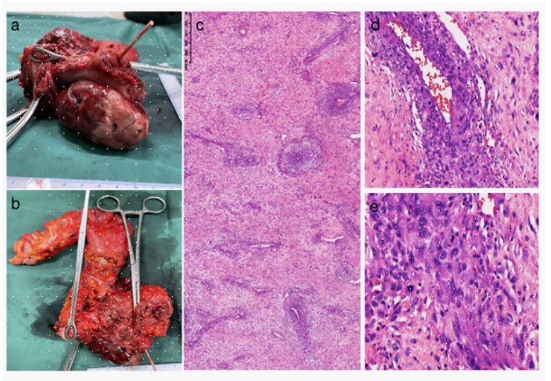
Histological features and histopathological specimen: Patient had surgeried the masses were resected and sent for histopatho logical examination (Figures 2a and b). Under the microscope, the tumor shows diffuse infiltrative growth, with significant atypia and pleomorphism of tumor cells. It surrounds blood vessels, with interstitial edema accompanied by inflammatory cell infiltration. Intravascular tumor thrombi are visible, and no clear nerve invasion is observed (Figures 2c,d and e). The tumor involves the bladder and rectum. IC showed similar pattern (Figure 2). Thus, the final diagnosis was PRS. The patient was advised regular follow up and not given any further RT or chemotherapy.
Discussion
Some patients suffering cancer may develop a second malignancy caused by primary anticancer treatment. PRS is a complications of Radiotherapy (RT) that occur within a previously irradiated field after several years of latency. This type of sarcomas are rare, and the latent period of at least 5 years and occur even after 60 years of radiotherapy. For cervical and endometrial cancers requiring radiation therapy, the PRS datum are limited. Jahanvi Gandhi et al. reported a rare case of post radiation Malignant Peripheral Nerve Sheath Tumor (MPNST) of pelvic following radiotherapy for endometrium carcinoma. Siew-Yen Lai et al. reported the first documented case of UCS diagnosed in a 72-year-old lady after 11 years of Concurrent Chemoradiotherapy (CCRT) for cervical cancer stage 1B2. Radical resection remains the main method of treatment in radiotherapy-induced sarcomas. N.S. Hadke et al. showed a 45-year-old woman developed malignant fibrous histiocytoma after radiotherapy of cervical cancer stage IIB. These patients radiotherapy dose is between 62-90Gy. The threshold dose for radiotherapy -induced tumors is not known, though a dosage of 40-60Gy is reported [7,8]. Epidemiological data concerning irradiation-induced sarcoma risk showed an average relative risk of 1.42 at 1 Sv (unit of dose equivalent, for gamma irradiation 1Sv=1Gy). Doxorubicin is the main postoperative drug treatment, but the effective rate is very low, the quality of life of these patients is seriously decreased, and the survival time is short. The usual dose of radiotherapy for cervical and endometrial cancer is 80-90Gy [9,10]. Whether the radiotherapy dose should be reduced as much as possible without affecting the radiotherapy effect of the primary tumor, so as to reduce the risk of sarcoma after radiotherapy. Radiotherapy is the main cause of post-radiotherapy sarcoma, and its pathogenesis may be genomic instability caused by DNA damage. The case reported in this study manifested three years after radiotherapy for cervical cancer, a markedly shortened timeframe compared to previously documented occurrences of post-radiotherapy sarcomas. Given the unique clinical trajectory of this patient, who sequentially experienced cervical cancer and endometrial cancer, underwent both radiotherapy and chemotherapy, it is plausible that the cumulative impact of these treatments may have significantly compromised genomic stability, leading to an earlier onset of the disease. Therefore, we need to pay more attention to how to control the scope and dose of radiotherapy, prolong the survival of patients, and reduce the occurrence of PRS.
A comprehensive review of literature pertaining to postradiotherapy sarcomas reveals a predominant incidence in tumors of the head and neck, bone, and breast, with scarce documentation in the context of gynecological malignancies. The limited reports on post-radiotherapy sarcomas in gynecological tumors may be attributed to variations in radiotherapeutic protocols, encompassing factors such as the scope and dosage of radiation administered [11,12]. This study reports on a patient who sequentially developed endometrial cancer and cervical cancer. The patient did not undergo carcinogenic gene testing, and the genetic background remains unclear. Additionally, the patient received a high dose of radiotherapy, leading to the rapid occurrence of post-radiation sarcoma. Microscopic examination revealed characteristics predominantly composed of smooth muscle components and mesenchymal tissue origin. This is considered a mixed type of post-radiation sarcoma. No similar cases have been identified in the literature, highlighting its clinical significance. The patient succumbed to the disease six months postoperatively, underscoring its highly malignant nature. With the aging population, the incidence of tumors is increasing. For tumor patients requiring radiotherapy, careful consideration should be given to treatment plans, especially in controlling radiation dosage. Future research should prioritize investigations into related aspects, given the potential implications for the growing population of elderly individuals and the rising prevalence of tumors.
Declarations
Data sharing statement: All inquiries can be directed to the corresponding authors.
Ethics statement and consent for publication: Institutional approval was not required to publish the case details. The patient provided written informed consent to the publication of this case report and any accompanying images.
Disclosure: The authors have no conflict of interest.
Acknowledgments: The authors thank to the patient and her family for their support of this stud
Additional information
Funding: This study has no funding.
References
- Weatherby RP, Dahlin DC, Ivins JC. Postradiation sarcoma of bone: review of 78 Mayo Clinic cases. Mayo Clinic Proceedings. 1981 May 56(5): 294-306. DOI: 10.2147/OTT.S123456
- Arora P, Wadhwa R, Khurana N, Jain S, Hadke NS. Secondary malignant fibrous histiocytoma of thigh with surface ulceration following radiotherapy for carcinoma cervix. Archives of Gynecology and Obstetrics. 2011 Mar 283 Suppl 1: 79-82. DOI: 10.1007/ s00404-010-1699-30.
- des Guetz G, Chapelier A, Mosseri V, et al. Post irradiation sarcoma: Clinicopathologic features and role of chemotherapy in the treatment strategy. Sarcoma. 2009; 2009: 764379. DOI: 10.1155/2009/764379.
- Almohsen SS, Alnuaim H, Salim AA, Arabi H. Pelvic Radiation-Induced Sarcoma With Rhabdomyoblastic Differentiation Following Treatment of Cervical Cancer. Cureus. 2021; 13(6): e15428. DOI: 10.7759/cureus.15428.
- Patel SR. Radiation-induced sarcoma. Current Treatment Options in Oncology. 2000; 1(3): 258-61. DOI: 10.1007/s11864-000-0037-6.
- Bonito FJP, Cerejeira DA, Dahlstedt-Ferreira C, Coelho HO, Rosas R. Radiation-induced angiosarcoma of the breast: A review. The Breast Journal. 2020; 26(3): 458-463. DOI: 10.1111/tbj.13504.
- Amendola BE, Amendola MA, McClatchey KD. Radiation-associated sarcoma: A review of 23 patients with postradiation sarcoma over a 50-year period. Am J Clin Oncol. 1989; 12(5): 411-415. DOI: 10.2147/OTT.S123462
- Taghian A, de Vathaire F, Terrier P, Le M, Auquier A, et al. Longterm risk of sarcoma following radiation treatment for breast cancer. Int J Radiat Oncol Biol Phys. 1991; 21(2): 361-367. DOI: 10.2147/OTT.S123463
- Akila NV, Sushil B, Demanes DJ, et al. American Brachytherapy Society consensus guidelines for locally advanced carcinoma of the cervix. Part II High-dose-rate brachytherapy. Brachytherapy. 2012; 11(1): 47-52. DOI: 10.2147/OTT.S123464
- Abu-Rustum NR, Yashar CM, Bean S, et al. NCCN clinical practice guidelines in oncology: NCCN guidelines for cervical cancer. Version 1. 2020 [EB/OL] 2020; 2020. DOI: 10.2147/OTT.S123465
- Liao Y-H, Lou P-J, et al. Radiation-induced sarcoma of head and neck: Clinical characteristics and molecular signatures. Head & Neck. 2023; 45(3): 638-646. DOI: 10.1002/hed.27279.
- Alamer OB, El Ahmadieh TY, et al. Primary and radiation-induced skull base osteosarcoma: A systematic review of clinical features and treatment outcomes. Journal of Neuro-Oncology. 2021; 153(2): 183-202. DOI: 10.2147/OTT.S123467.