Open Journal of Clinical and
Medical Images
Clinical Image - Open Access, Volume 5
Gangrenous keloid of ear lobe: A clinical image
Sanjay C Babar1; Ashwini Supekar2*
Department of Shalyatantra, Dr DY Patil College of Ayurved and Research Centre, Dr DY Patil Vidyapeeth (Deemed to be University) Pimpri, India.
*Corresponding Author: Ashwini Supekar
Department of Shalyatantra, Dr.D Y Patil College of
Ayurved & Research Centre, Dr DY Patil Vidyapeeth
(Deemed to be University) Pimpri,
Pune-18, Maharashtra, India.
Email: ashwinisupekar1997@gmail.com
Received : Dec 17, 2024
Accepted : Jan 10, 2025
Published : Jan 17, 2025
Archived : www.jclinmedimages.org
Copyright : © Supekar A (2025).
Citation: Babar SC, Supekar A. Gangrenous keloid of ear lobe: A clinical image. Open J Clin Med Images. 2025; 5(1): 1200.
Description
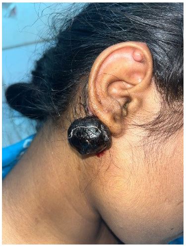
A keloid is an abnormal proliferation of scar tissue that forms at the site of cutaneous injury. Keloids are benign dermal fibro proliferative tumors with no malignant potential. A 17-year-old female patient came with complaint of Pedunculated necrosed mass over right ear lobe since 2 days of approx. size 5 x 5 cm. She gave a history of ear piercing 6 months back. Since then, the swelling started to grow. Since last 2 days the swelling started to turn dark and patient had severe pain and itching over it. The swelling was firm & tender on examination. The pedunculated keloid, being attached to the skin by a narrow base, which restricted blood flow to the tissue, leading to tissue death. As the keloid grows larger, it exerted pressure on the blood vessels, reducing oxygen and nutrient supply to the tissue. So, Clinical diagnosis of Gangrenous Keloid was made. The patient must be having keloid tendency because she had developed a keloid on the superior helix of same ear also after piercing. The swelling was then excised under local anesthesia & primary suturing was done.